
Anatomy of Mussels
- Pearl Farming Training Team
- Jul 12, 2021
- 1 min read
Updated: Jul 24, 2021


Mussel is the common name used for members of several families of bivalve mollusks, from saltwater and freshwater habitats. These groups have in common a shell whose outline is elongated and asymmetrical compared with other edible clams, which are often more or less rounded or oval.
The common name "mussel" is also used for many freshwater bivalves, including the freshwater pearl mussels. Freshwater mussel species inhabit lakes, ponds, rivers, creeks, canals, and they are classified in a different subclass of bivalves, despite some very superficial similarities in appearance.
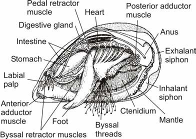
The mussel's external shell is composed of two hinged halves or "valves". The valves are joined together on the outside by a ligament, and are closed when necessary by strong internal muscles (anterior and posterior adductor muscles). Mussel shells carry out a variety of functions, including support for soft tissues, protection from predators and protection against desiccation.
The shell has three layers. In the pearly mussels there is an inner iridescent layer of nacre (mother-of-pearl) composed of calcium carbonate, which is continuously secreted by the mantle; the prismatic layer, a middle layer of chalky white crystals of calcium carbonate in a protein matrix; and the periostracum, an outer pigmented layer resembling a skin. The periostracum is composed of a protein called conchin, and its function is to protect the prismatic layer from abrasion and dissolution by acids (especially important in freshwater forms where the decay of leaf materials produces acids).
Source: Wikipedia